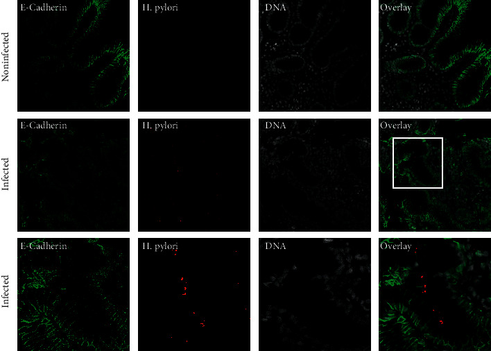
Figure 1.

Detection of H. pylori in gastric biopsy specimens. Sections of gastric glands obtained from H. pylori-positive patients fluorescently labeled for the epithelial marker E-cadherin (green), H. pylori (red), and nuclear DNA using DAPI (white). Most of the gastric glands are heavily infected with H. pylori.
