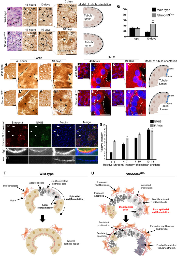
Figure 4.
Actomyosin signaling during epithelial kidney repair is Shroom3 dependent. (A)–(F) Representative images of H&E demonstrating altered tubular epithelial morphology, (A) and (D) dotted line and arrowheads, and persistent tubular epithelial injury by Kim1 IHC expression (B), (C), (E), and (F) dotted line and arrowheads, 10 days after IRI injury. (G) Quantitative analysis demonstrating Kim1 expression in the cortical tubular epithelium in WT and Shroom3Gt/+ mutants 10 days after IRI. (H)–(O) Representative low- and high-power images of immunostaining for F-actin and phosphorylation of myosin light chain (pMLC) in WT and Shroom3Gt/+ mutants 48 hours and 10 days after IRI. Then 10 days after kidney injury, F-actin and pMLC protein expression is apically localized in WT tubular epithelium but not in Shroom3Gt/+ mutants (dotted line and arrowheads). (P)–(R) Representative low- (P) and high-power (Q), (R) images of the MDCK apical cell membrane after immunolabeling for Shroom3, nonmuscle myosin IIB (NMIIB), and F-actin. (Q) High expressing Shroom3 cells exhibit robust immunofluorescent signal in the apical membrane (*). (R) Low-expressing Shroom3 cells exhibit a reduced apical NMIIB and F-actin expression (arrowheads). (S) Quantitation of pixel density of MDCK cells immunolabeled for Shroom3, NMIIB, and F-actin. Low-expressing Shroom3 cells express lower levels of NMIIB and F-actin compared with the higher expressing Shroom3 MDCK cells. *P≤0.0001. Scale bars = 40 µm (A)–(O), insets = 10 µm. WT = wild-type.

