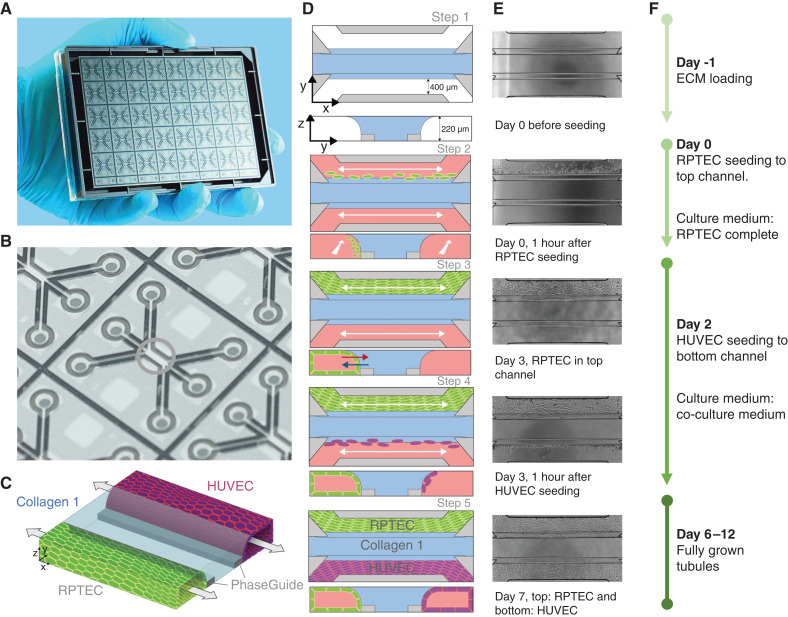
Figure 1.
Overview of the seeding method of the renal proximal tubule epithelial cells (RPTEC)/human umbilical vein endothelial cells (HUVEC) coculture in the OrganoPlate 3-lane. (A) Photograph of the bottom of the culture platform showing 40 microfluidic channel networks underneath a 384-well plate. (B) Zoom-in on a single microfluidic channel network comprising three channels that join in the center (green circle). (C) Three-dimensional artist impression of the center of a chip, where two tubules are cultured in the two lateral channels (green and purple) along an extracellular matrix (ECM) gel in the middle channel (light blue). Two phaseguides (gray bars) define the positioning of the ECM gel leading to the three-lane stratified profile. (D) Artist impression of the horizontal projection and vertical cross-section. (E) Associated phase-contrast images. (F) Timeline for setting up the coculture.

