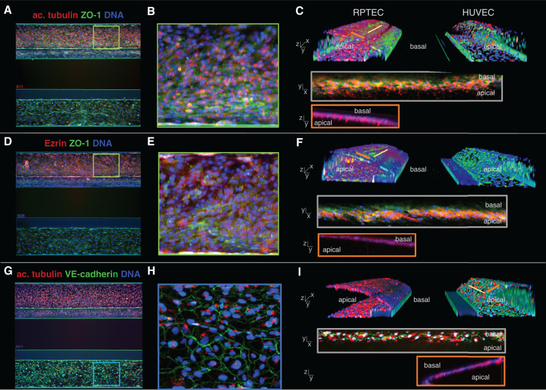
Figure 2.
Marker expression of the kidney model shows polarized epithelium and endothelium. (A, D, and G) Max z-projections of the coculture with the RPTEC tubule in the top channel and the HUVEC vessel in the bottom channel. (B, E, and H) Zoom of the z-projections in (A), (D), and (G). (C, F, and I) three-dimensional reconstructions showing a view into the lumen of the tubules. (A–C) Primary cilia were visualized by acetylated tubulin staining (red), present on the apical side of the RPTEC tubule. Tight junction protein ZO-1 (green) was present in both cell types. (D–F) Epithelial marker and brush border protein Ezrin (red) was exclusively present on the apical side of the RPTEC tubule. (G–I) Endothelial tight junction protein VE-cadherin (green) was expressed by the HUVEC vessel at the cell border, and primary cilia located on the apical side of the membrane were stained using acetylated tubulin.

