Figure 4.
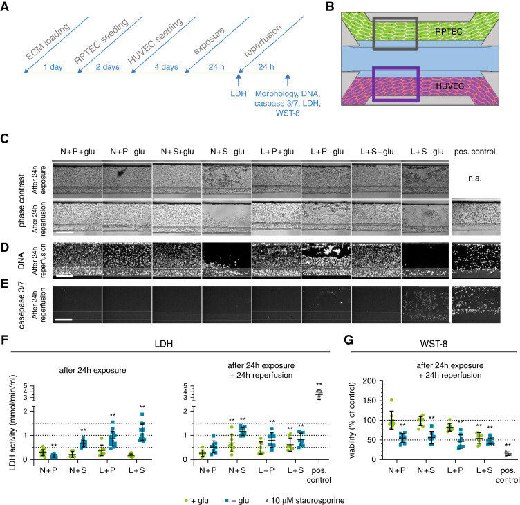
Ischemic conditions lead to AKI in the proximal tubule. Ischemia was modeled on the OrganoPlate coculture through a combination of low oxygen (L), static incubation (S), and glucose- and nutrient-poor medium (-glu) for 24 hours, followed by a 24-hour reperfusion in normoxia (N), perfusion on the rocker (P), and in glucose- and nutrient-rich medium (+glu). (A) Timeline of the experiment. (B) Region of the RPTEC tubule (gray square) that is used for the images shown in (C–E). (C) Representative phase-contrast images after 24-hour exposure (top) and subsequent 24-hour reperfusion (bottom). Different ischemia inducing conditions were tested (columns) and compared with the normal condition N+P+glu. n.a., not available. (D) DNA staining after 24-hour reperfusion. (E) Caspase-3/7 staining after 24-hour reperfusion. Scale bar, 200 µm. (F) LDH release in the medium was measured after 24-hour exposure (left) and 24-hour exposure plus 24-hour reperfusion (right), respectively. (G) WST-8 viability relative to the normal condition N+P+glu was assessed after 24-hour reperfusion. Staurosporine (10 µM) was included as a positive control. Error bars represent standard deviation. One-way ANOVA compares the conditions to the N+P+glu control condition, **P<0.01, n=8–16 chips per condition.

