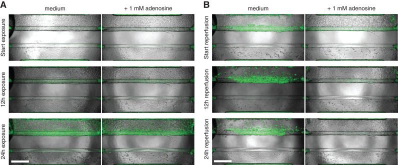
Figure 6.
Real-time caspase-3/7 activation and phase-contrast imaging shows the protective effect of adenosine upon ischemic exposure. (A) Cultures, coincubated with and without 1-mM adenosine, were exposed to ischemic conditions (L+S-glu) for 24 hours, and caspase-3/7 activity was monitored over time. (B) Medium was refreshed to standard culture medium, and cocultures were reperfused under normal conditions (N+P+glu) for 24 hours. Green: activated caspase-3/7. Scale bar: 500 µm. Representative images of n=3 chips per condition. A corresponding time-lapse movie can be viewed in the Supplemental Video.

