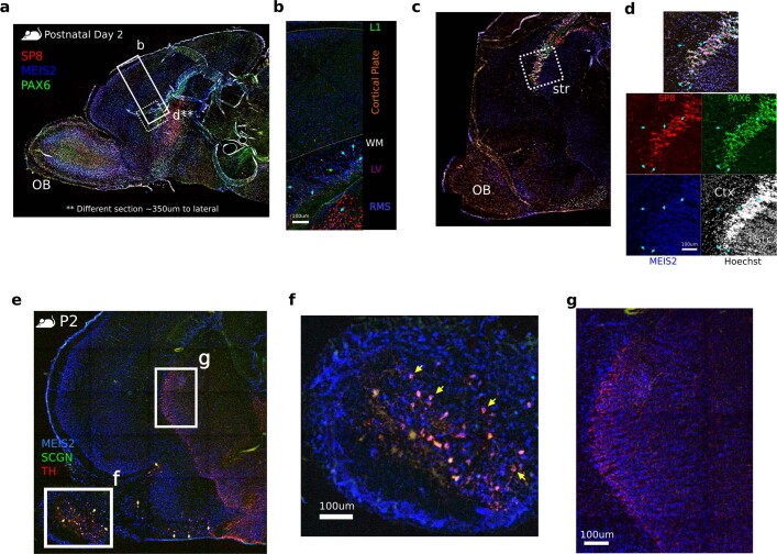
Extended Data Fig. 14. Distribution of LGE_MEIS2/PAX6-derived cells in postnatal mouse.
a. Mouse sagittal section showing b and approximate c magnification locations. b. LGE_MEIS2/PAX6 cells in deep white matter (cyan arrows) c. Lateral sagittal section d. Panel showing SP8+/MEIS2+/PAX6+ cells in remainder of dLGE, likely homologous to dLGE chains in Arc. e. Lateral sagittal section of mouse postnatal day 2. f. MEIS2+/SCGN+/TH+ periglomerular cells in lateral OB. g. Striatum shown with dense TH+ projection fibers and synapses, but no MEIS2+/SCGN+/TH+ cell bodies.

