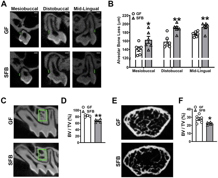
Figure 2. SFB has catabolic effects on alveolar bone.
A-B. Alveolar bone loss was measured by calculating the linear distance between the cementoenamel junction (CEJ) and alveolar bone crest (ABC) at the maxillary first molar in reconstructed micro-CT images. A. Representative micro-CT images displaying CEJ-ABC linear distance (green line) at the mesiobuccal line angle, distobuccal line angle, and mid-lingual aspect of the maxillary first molar; scale bar (white line) = 200μm. B. Quantitative measures of CEJ-ABC linear distance at the mesiobuccal line angle, distobuccal line angle, and mid-lingual aspect of the maxillary first molar (n=6/gp). C. Representative micro-CT images of trabecular bone in the maxillary first molar trifurcation, region of interest outlined (green box). D. Micro-CT quantitative analysis of trabecular bone volume per tissue volume (BV/TV) in the maxillary first molar furcal alveolar bone (n=4/gp). E. Representative micro-CT reconstructed cross-sectional images of trabecular bone in the distal femur, extending 50μm proximally from where analysis began. F. Micro-CT quantitative analysis of BV/TV in the distal femur (n=6/gp). Unpaired t-test; data are presented as mean ± SEM; *p<0.05, **p<0.01.

