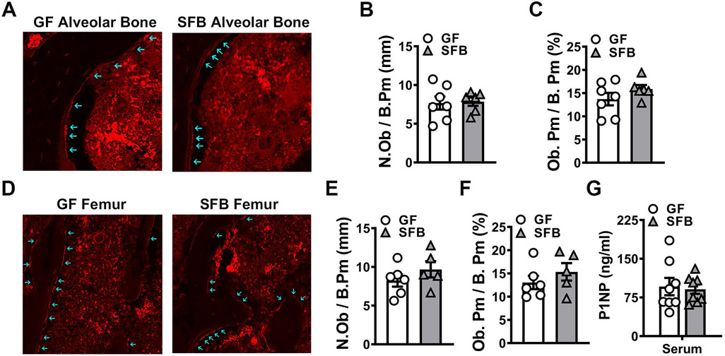
Figure 4. SFB colonization does not alter osteoblastogenesis.
A-C. Immunofluorescent analysis of osteoblasts in the maxillary first molar furcal alveolar bone (n=6-7/gp). Osterix+ cuboidal cells lining bone were designated as osteoblasts (red, osterix – Rhodamine). A. Representative images of osterix+ osteoblasts lining the maxillary first molar furcal alveolar bone; arrows indicate labeled osteoblasts (200X). B. N.Ob/B.Pm = osteoblast number per bone perimeter. C. Ob.Pm/B.Pm = osteoblast perimeter per bone perimeter. D-F. Immunofluorescent analysis of osteoblasts in the secondary spongiosa of distal femur (n=5-6/gp). Osterix+ cuboidal cells lining bone were designated as osteoblasts (red, osterix – Rhodamine). D. Representative images of osterix+ osteoblasts lining the secondary spongiosa trabecular bone of distal femur; arrows indicate labeled osteoblasts (200X). E. N.Ob/B.Pm. F. Ob.Pm/B.Pm. G. Serum ELISA analysis of P1NP levels (n=8/gp). Unpaired t-test; data are reported as mean ± SEM.

