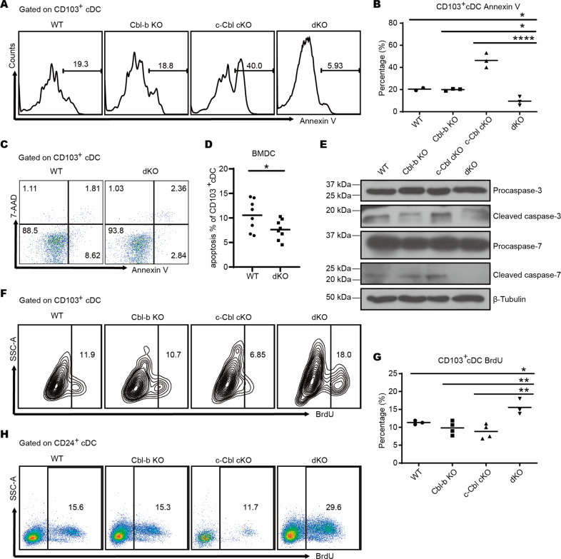
Fig. 3. Ablation of Cbl-b and c-Cbl decreases the apoptosis and increases the proliferation of cDC1s.
A Leukocytes isolated from livers were stained with surface markers, subjected to Annexin V staining, and assessed with flow cytometry. n = 3 per group. B The percentage of Annexin V+ CD103+ cDC1s in liver. C, D Bone marrow cells of WT and dKO mice were cultured with 20 ng/ml Flt3-L for 7 days and 20 ng/ml Flt3-L plus 2 ng/ml GM-CSF for another 2 days. The percentage of apoptotic cells was determined by flow cytometry. n = 8 per group. E CD103+ cDCs (MHC-II+ CD11c+) differentiated in (C) were sorted and the protein expression of caspase-3 and caspase-7 was measured by Western blot. F Mice were injected with 1 mg BrdU 24 h and 6 h prior to sacrifice, respectively. Proliferation of liver cDCs was quantified using flow cytometry. n = 4 per group. G The percentage of BrdU+ CD103+ cDC1s in the liver. H Bone marrow cells of WT and dKO mice were cultured as in (C) followed by 10 µM BrdU incubation for 6 h and quantification via flow cytometry. *p < 0.05, **p < 0.01, ****p < 0.0001, ns, no significance, One-Way ANOVA comparisons for B and G, unpaired Student’s t test for (D). p < 0.05 was considered statistically significant.

