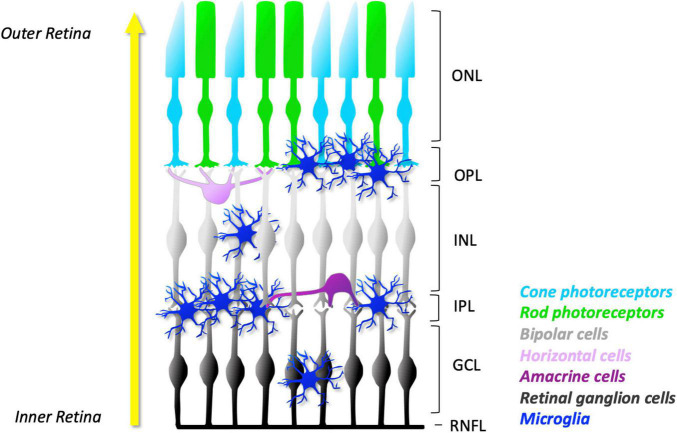
FIGURE 1.
Schematic representation of microglial distribution in layers of normal mature retina. As development progresses, microglia migrate from the GCL and IPL towards the outer layers of the retina (yellow arrow). In the young retina, the number of microglia in their ramified appearance is found predominantly in the synaptic layers: the OPL and IPL. They can also be found in lesser numbers in the GCL and INL. However, no microglia are found in the ONL, a specialised microglial exclusion zone. The concentration of microglial processes in the plexiform layers facilitates frequent and dynamic contact with neuronal dendrites, axons, and synapses. The ramified morphology allows microglia to constantly extend and retract their processes. Together, these features facilitate constant surveillance of the surrounding microenvironment. ONL, outer nuclear layer; OPL, outer plexiform layer; INL, inner nuclear layer; IPL, inner plexiform layer; GCL, ganglion cell layer; RNFL, retinal nerve fibre layer.

