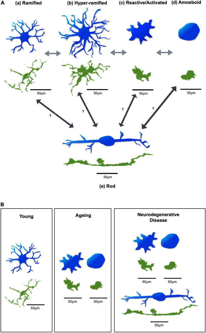
FIGURE 2.
Diagram of microglial morphologies. (A) Microglial morphology varies based on activation state, in response to changing environmental conditions. Arrows demonstrate possible transitions between states. These transitions can occur in both directions, allowing microglia to switch back and forth between activation states. (a) Microglia under resting physiological conditions have a ramified appearance. (b) Disruption to environmental homeostasis leads them to elongate their processes and transiently exhibit a hyper-ramified state. (c) In response to considerable environmental damage, they rapidly adopt an activated morphology, with an increased soma size, and thicker, shorter processes. (d) Substantial insult leads them to adopt an amoeboid appearance, with complete retraction of processes, allowing directed motility and phagocytosis of target material. (e) Rod microglia, which are specifically associated with retinal neurodegeneration, exhibit a uniquely narrow and elongated morphology, with few processes. It is unclear which microglial activation state(s) give(s) rise to rod microglia. Figure based on Holloway (Holloway et al., 2019). (B) Different predominant morphologies and activation states of microglia may be found in the young, ageing, and neurodegenerative retina. Generally, ramified microglia may be predominantly found in the young retina. In the ageing retina, the reactive/activated and amoeboid microglia are predominantly found. In cases of retinal neurodegenerative disease, reactive/activated, amoeboid, and rod microglia are predominantly found. Blue microglia are illustrative representations to demonstrate morphological changes. Green microglia are from whole-mount retinal images of Iba-1 stained C57BL6 mice, obtained from the Cordeiro laboratory. Scale bar = 50 μm.

