
Fig. 4.
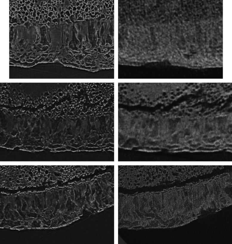
A still image of Video 1 showing the annual layers are detectable in the movies in the left column and at the one on the bottom in the right column: Scroll-through 160 cross-sectional virtual histology slices, prepared by the software VGStudio MAX, of the human tooth obtained at the ANATOMIX beamline (images in the left column) and corresponding local scans by means of SkyScan 2214 without phase retrieval (right column, top row), of Exciscope with a priori phase retrieval (right column, middle row), and of Xradia 620 Versa without phase retrieval (right column, bottom row). The width of each box equals about (Video 1, MP4, 11639 KB [URL: https://doi.org/10.1117/1.JMI.9.3.031507.1]).