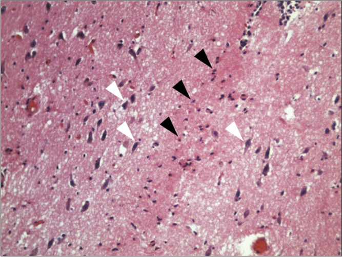
Fig. 3.
Histological view of a horizontal section of the interthalamic adhesion (zoom ×20), in which the absence of gray commissure it is notable, but glial cells are distinguished (black triangles), and laterally, soma of pyramidal neurons (white triangles), with anterior to posterior soma orientation in hematoxylin and eosin stain.

