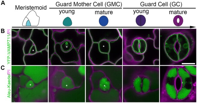
Figure 1.
Vacuole development and distribution in stomatal lineage cells of Arabidopsis cotyledon epidermis. A, Schematic diagram showing color-coded models of stomatal lineage cells in five developmental stages: Meristemoid, cyan; young GMC, green; mature guard mother cell, blue; YGC, indigo; and mature GC, purple. B and C, Dynamics and subcellular localization of the two distinct organelle markers in the five corresponding stages of stomatal lineage cells, including the tonoplast marker YFP-VAMP711 (B), and the vacuolar lumen marker Aleu-Kaede (C). Asterisks indicate the corresponding lineage cells in each stage. Cell walls stained with PI (magenta). Scale bar, 10 μm for all panels.

