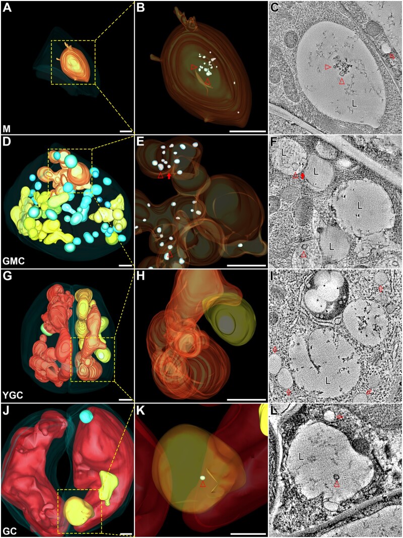
Figure 3.
Swollen ILVs are present in the large vacuoles of the stomatal lineage cells. The indicated square boxes in each whole-cell ET model of M (A), GMC (D), YGC (G), GC (J) were further zoomed-in for ET analysis with their corresponding enlarged models shown in (B), (E), (H), (K), respectively. Their representative tomographic slices were shown in (C), (F), (I), (L), respectively. Semi-transparent models of large vacuoles display internal details. Small vacuoles were omitted in GMC to avoid unexpected block (E). Arrowheads indicate examples of swollen ILVs in 3D ET models (B, E, and K) and their corresponding tomographic slices (C, F, and L). Note that multiple large vacuoles in GMC are interconnected (E and F). The closed arrow indicates a direct membrane connection between the two large vacuoles (F). The open arrows indicate examples of the lipid droplets (I). M, meristemoid; L, large vacuole. Scale bars, 1,000 nm for all the figure parts.

