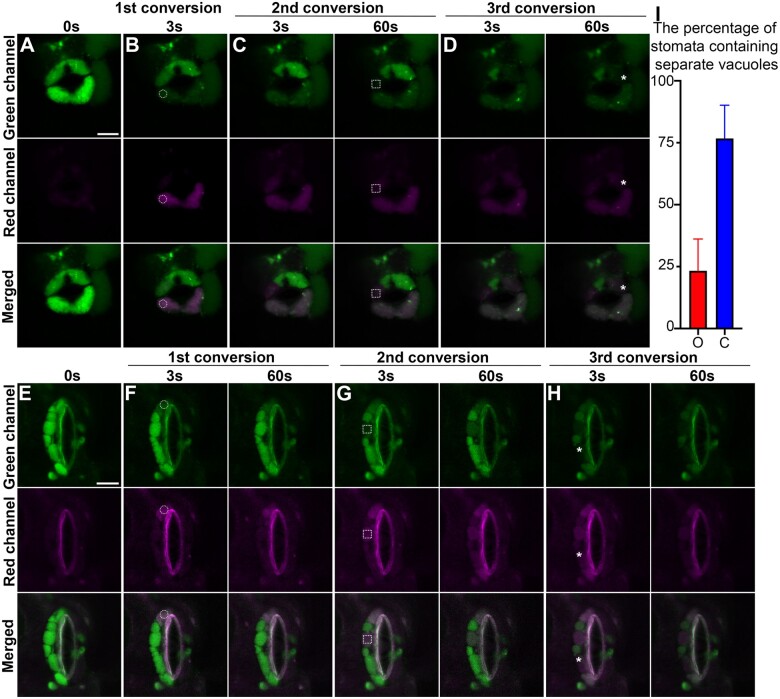
Figure 5.
In vivo physical relationship of vacuoles in opening and closed stomata from Arabidopsis cotyledon epidermis. Opening and closed stomata from transgenic Arabidopsis plant, which expresses the photoconvertible vacuole-localized marker Aleu-Kaede, were subjected to photo-activation and conversion experiments sequentially at three time points as indicated, followed by confocal imaging at respective channels. The circles in (B and F), the square boxes in (C and G), and the asterisks in (D and H) indicate the positions of each photo-activation, respectively. The green channel (top) shows the original Aleu-Kaede signals whereas the Red channel (middle) exhibits the activated/converted signals after photo-activation. Scale bars, 10 μm for all figure parts. A and E, Control prior to photo-activation and conversion, showing the vacuoles with luminal green signals in the two GCs. B and F, Confocal imaging at 3 s (B and F) and 60s (F) post first conversion. The white circles indicate the regions of photo-activation. C and G, Confocal imaging at 3 s and 60 s post second conversion, respectively. The white square boxes indicate the regions of photo-activation. D and H, Confocal imaging at 3 s and 60 s post third conversion, respectively. The asterisks indicate the regions of photo-activation. I, Statistical analysis on the percentage of stomata containing separate vacuoles. O, opening stomata; C, closed stomata. Data are shown as mean ± sd of three repeats. n = 20.

