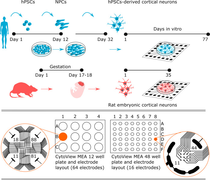
Fig. 1.
Experimental setup for following the functional development of human and rat networks on two different MEA well plate types. After human pluripotent stem cells (hPSCs) differentiated into cortical neurons, they were plated on CytoView MEA 12 and CytoView MEA 48 well plates. Rat embryonic cortical cells were plated on the same MEA well plate types in parallel. Regular timepoint recordings were obtained in CytoView MEA 12, which has 64 electrodes per well, and pharmacological experiments were performed on CytoView MEA 48, which has 16 electrodes per well. Measurement days in MEA are referred to as days in vitro (DIV). Pharma recordings were obtained at DIV 29 and DIV 22, when peak activity is commonly observed for in vitro human and rat neuronal networks, respectively.

