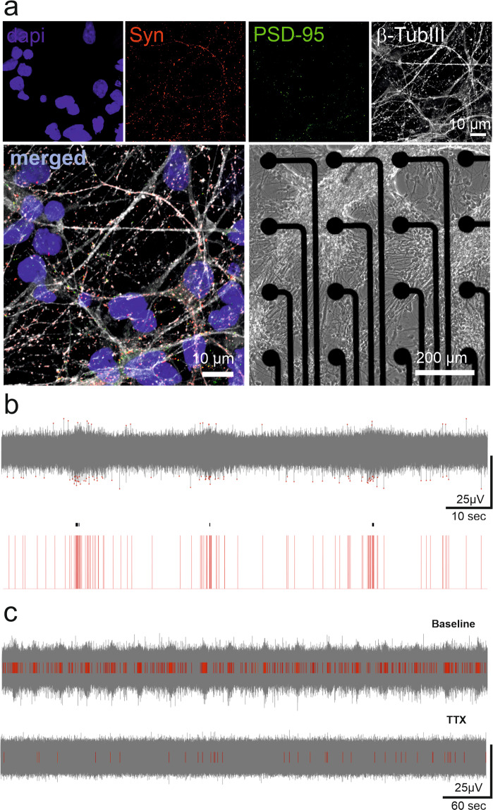
Fig. 3.
Validation of the MEA recordings of neuronal networks and signal analysis. (a) The quality of hPSC-derived neural cultures was verified with immunocytochemical staining of pre- and postsynaptic markers (synaptophysin in red and PSD-95 in green) overlaid with neuron-specific b-tubulin staining (white). DAPI stains cell nuclei. A phase contrast image of hPSC-derived neurons on the MEA is used to illustrate the formation of networks over the electrode area. (b) The performance of spike detection in recognizing the typical low-amplitude signal of hPSC-derived networks was confirmed. The detected spikes are illustrated with red dots (top) and red bars (bottom). A single channel burst detection algorithm successfully detected quasiperiodic burst activity, which is commonly observed in mature neuronal cultures. The detected bursts are labeled with black lines on the red spike bars (bottom). (c) The MEA signal was associated with action potentials, as it was blocked by the voltage-gated sodium channel antagonist tetrodotoxin (TTX). The number of detected spikes (red bars) decreased considerably (bottom) compared to the baseline recording (top).

