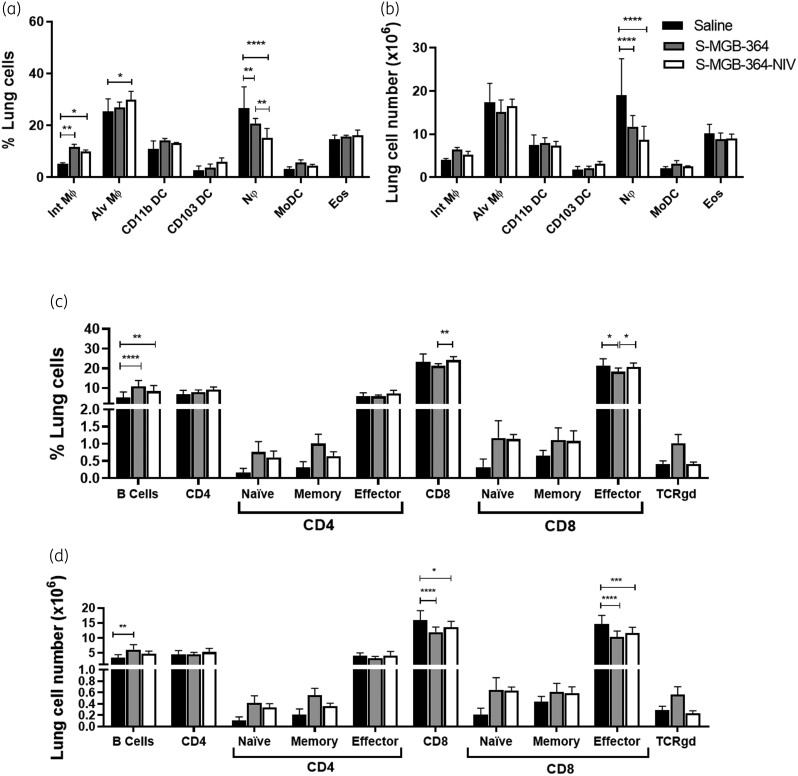
Figure 5.
Increased lung macrophage and B cell recruitment and decreased CD8+ T cells and neutrophil recruitment in S-MGB-364- and S-MGB-364-NIV-treated mice following Mtb HN878 infection. C57BL/6 mice (n = 6 per group) were infected with 100 cfu of Mtb HN878 strain via intranasal challenge. At 1, 2, 3 and 4 weeks post infection, mice were intranasally treated with 10 mg/kg of S-MGB-364, S-MGB-364-NIV, and saline. Mice were sacrificed at 5 weeks post-infection and cellular infiltration was analysed from lung cell suspensions by flow cytometry. (a) Percentage and (b) total cell numbers of myeloid populations, (c) percentage and (d) total cell numbers of lymphoid populations from lung single cell suspensions of mice treated with S-MGB-364, S-MGB-364-NIV or saline. Alveolar macrophages (Alv MΦ) = CD64+SiglecF+CD11c+; interstitial macrophages (Int. MΦ) = MERTK+CD64+CD11c−SiglecF−; CD103 dendritic cells (DC) = MHCII+CD11c+CD103+CD11b−, CD11b DC = MHCII+CD11c+CD103−CD11b+; neutrophils (Nφ) = LY6G+CD11b+; monocyte-derived dendritic cells (MoDC) = CD64+ CD11b+CD11c+; eosinophils (Eos) = CD64−SiglecF+CD11b+; B cells = CD19+CD3−; CD8+ T cells = CD3+CD4−CD8+; CD4+ T cells = CD3+CD4+CD8−; naive T cells = CD62L+CD44−; memory T cells = CD62L+CD44+; effector T cells = CD62L−CD44+; TCRγδ Cells = TCRγδ+CD3+. Data shown as mean ± SEM. One-way ANOVA: *P < 0.05, **P < 0.01; ***P < 0.001, ****P < 0.0001.

