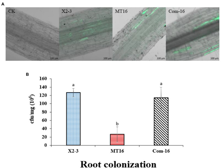
Figure 3.
Qualitative and quantitative analysis of wheat root colonization by Lysobacter capsici X2-3 cells. The roots were cultured in X2-3, MT16, and Com-16 for 3 days. (A) Confocal scanning laser microscopy images of the roots colonized by L. capsici. Wheat roots without gfp inoculation as a control. Wheat roots colonized with X2-3-gfp, MT16-gfp, and Com-16-gfp for 3 days. Bar = 100 μm. (B) Quantitative analysis of root colonization by wild-type L. capsici, the LC_GidA deletion mutant and the complemented strain. a, not significant compared to X2-3. b, significant difference compared to X2-3.

