FIGURE 8.

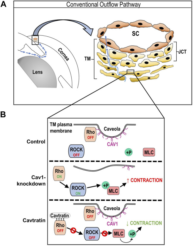
Schematic of hypothesized regulation of Rho/ROCK activity by Cav1 in TM cells. (A) Cartoon showing components of the irideocorneal angle, highlighting aqueous humor flow (blue arrows) into and through the conventional outflow pathway. A higher magnification illustration of the conventional outflow pathway, depicting the TM, juxtacanalicular tissue (JCT) and SC on the right. (B) Diagram demonstrating results of the present study, showing the status of Rho/ROCK activity and MLC phosphorylation in Cav1 competent, Cav1 deficient, and cavtratin-treated TM cells. For example, we propose that in the presence of Cav1, RhoA localizes to the plasma membrane, where it is sequestered and inhibited via physical interaction with the Cav1 scaffolding domain, leading to a low basal level of Rho/ROCK activity and MLC phosphorylation. In the absence of Cav1, we propose that RhoA is liberated and basal Rho/ROCK activity and MLC phosphorylation is elevated. Following treatment with the Cav1 scaffolding domain peptide, cavtratin, we hypothesize that RhoA is inhibited, leading to reduced ROCK activity and subsequent MLC dephosphorylation.
