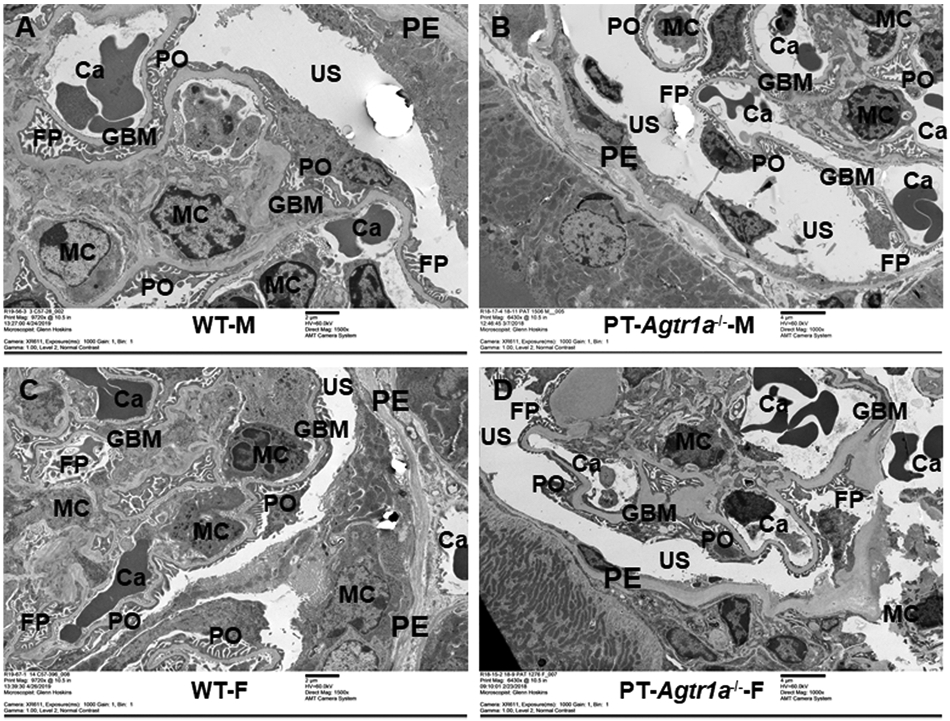
Figure 1. High resolution EM micrographs comparing the glomerular ultrastructures between male and female WT and proximal tubule-specific AT1 (AT1a) receptor-knockout mice, PT-Agtr1a−/− mice.

(A,C) High-resolution EM micrographs showing the ultrastructures of representative glomerulus (9720×) in adult male and female WT mice. (B,D) EM micrographs showing the ultrastructures of representative glomerulus (6430 or 9720×) in adult male and female PT-Agtr1a−/− mice, respectively. Glomerular ultrastructures including capillaries (Ca), epithelial (PE), mesangial cells (MCs), and POs were largely similar between male and female WT mice, or between male and female PT-Agtr1a−/− mice. Urinary space (US) of Bowman’s capsule, and foot processes (FP) looks similar between male WT (A) and PT-Agtr1a−/− mice (B), or between female WT (C) and PT-Agtr1a−/− mice (D). Likewise, GBMs were not different between male and female WT and PT-Agtr1a−/− mice. n=8 per group.
