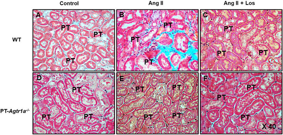
Figure 9. Comparisons of the proximal tubular and interstitial fibrotic responses, as determined by Masson’s trichrome staining, to Ang II-induced hypertension treated with or without losartan between male WT and PT-Agtr1a−/− mice.
(A) WT control mice. (B) WT mice with Ang II infusion. (C) WT mice with concurrent Ang II infusion and losartan treatment. (D) PT-Agtr1a−/− control mice. (E) PT-Agtr1a−/− mice with Ang II infusion. (F) PT-Agtr1a−/− mice with concurrent Ang II infusion and losartan treatment. Note that Ang II induced marked tubulointerstitial fibrotic response in WT mice (B), compared with control WT mice (A), and Ang II-induced tubulointerstitail injury response was completely blocked by concurrent losartan treatment (C). By contrast, Ang II did not significantly induce significant tubulointerstitial injury or fibrotic response in PT-Agtr1a−/− mice (E vs. D) with or without losartan treatment (F). PT, proximal tubule. Magnification: ×40.

