Figure 2.
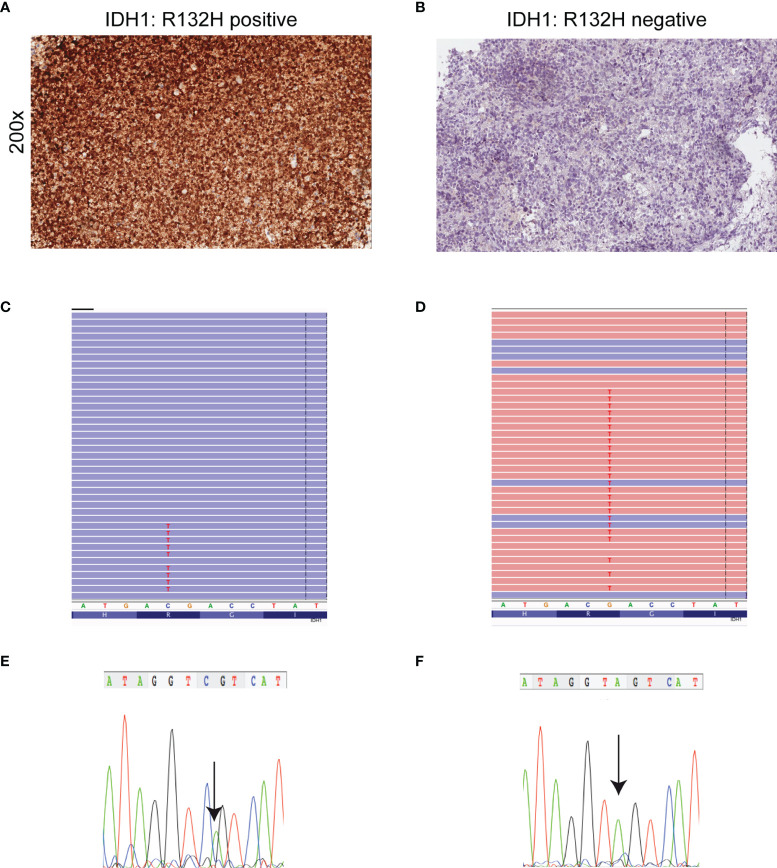
Detection of mutant IDH1 by immunohistochemistry, NGS and Sanger sequencing. (A) Astrocytoma cells are diffusely and strongly stained with anti-IDH1 (R132H) antibody (immunoperoxidase; original magnification 200x). (B) Corresponding IDH1 c.395G>A (p. R132H) mutation detected by NGS. Results were viewed in the Integrative Genomics Viewer (IGV). (C) The chromatogram showing representative sequencing results of IDH1 c.395G>A (p. R132H). (D) Absence of IDH1 (R132H) immunoreactivity in a IDH1- (R132S)-mutant astrocytoma. (E) IDH1 c.394C>A (p. R132S) mutation detected by NGS. Results were viewed in the IGV. (F) The chromatogram showing the representative sequencing results of IDH1 c.394C>A. Note that IDH1 is a negative-sense gene with respect to the genomic reference sequence. Thus, any nucleotide change is displayed as reverse complement. The arrow symbols on chromatogram indicate the place of mutation.

