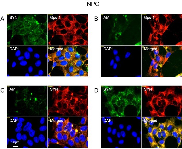
Fig. 7.

α-SYN, SYNfils and Gpc-1 co-localize in the cytoplasm of human NPC. Representative immunofluorescence images of confluent cultures of NPC. Staining was performed with mAb SYN (A, green), mAb AM (for HS-anMan, B, C, green), mAb SYNfil (D, green), pAb GPC-1 (A, B, red), pAb SYN (C, D, red) and DAPI (for nuclei, blue). Exposure time was the same in all cases. Bar, 20 μm.
