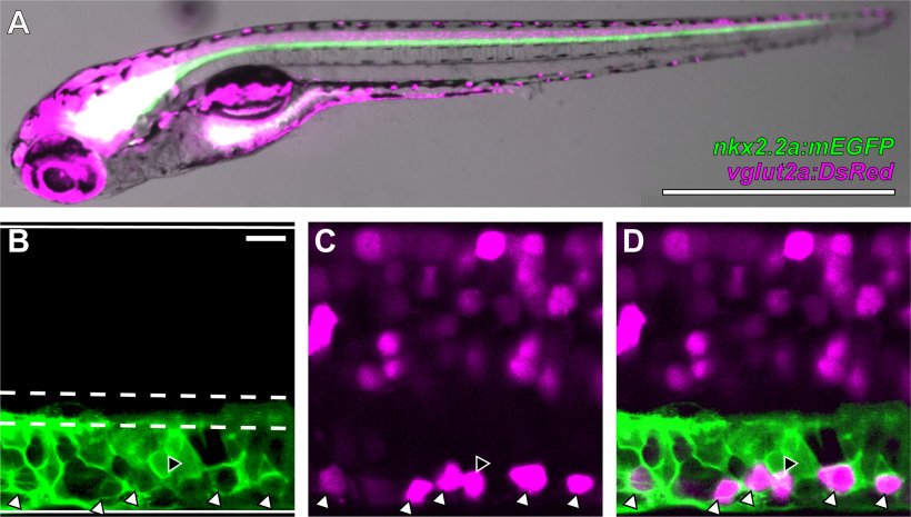
Figure 1.
V3-INs are identified by co-localization of vglut2a:DsRed and nkx2.2a:mEGFP transgene expression in neurons located in the ventromedial spinal cord. A, Whole-mount Tg(nkx2.2a:mEGFP)vu17;Tg(vglut2a:DsRed)nns9 double transgenic zebrafish larva. B–D, A representative single confocal optical section of the spinal cord exhibiting: (B) nkx2.2a:mEGFP expression, (C) vglut2a:DsRed expression, and (D) overlayed nkx2.2a:mEGFP and vglut2a:DsRed expression. The boundaries of the spinal cord and central canal are illustrated by solid and dashed white lines, respectively, in panel B. White triangles indicate neurons that co-express both transgenes and the black triangle indicates the only vglut2a:DsRed neuron ventral to the central canal that did not co-express nkx2.2a:mEGFP in the field of view. Scale bars: 1 mm (A) and 10 μm (B–D).

