Figure 3.
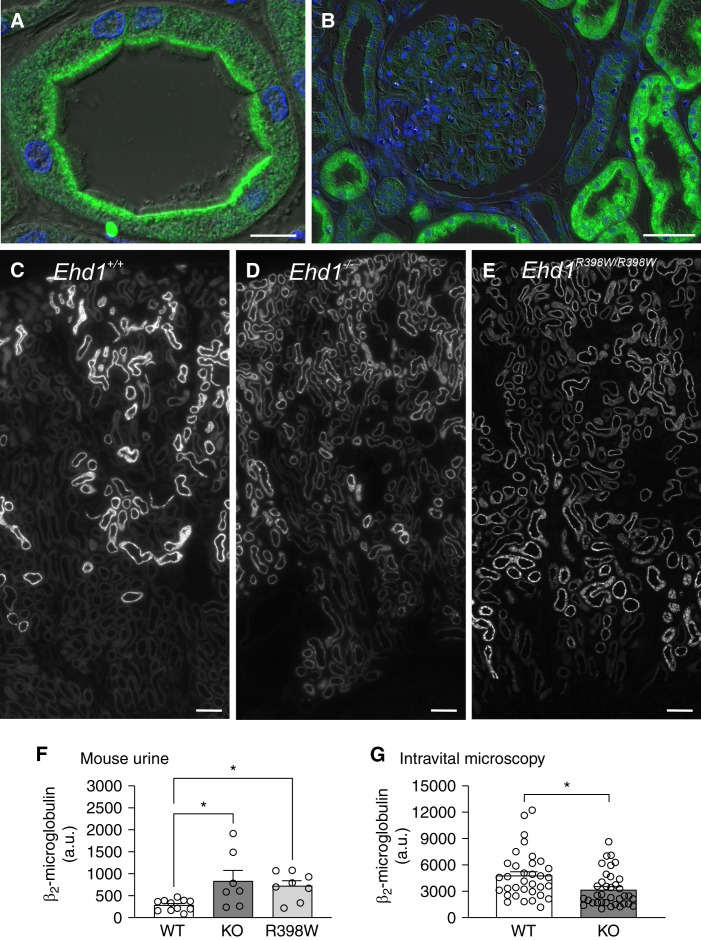
EHD1 localization and function in the kidney. (A) Localization of EHD1 (green) in the normal human proximal tubule. Please note strong EHD1 signal in the subapical compartment beneath the tubular lumen. Nuclear staining (blue); scale bar 10 µm. (B) Normal human kidney showing very little or absent expression of EHD1 (green) within the glomerulus and EHD1 prominence in adjacent proximal tubules. Nuclear staining (blue); scale bar 50 µm. (C–E) Kidney sections showing reabsorption of fluorescently labeled β2-microglobulin (white) (30 minutes after injection) in proximal tubules in a wildtype (C), knockout (D), and knockin mouse (E). Note, in wildtype (WT) mouse kidney (C) reabsorption of fluorescently labeled β2-microglobulin was observed mainly in early proximal tubules (S1 and S2 segments). In kidneys of homozygous Ehd1 knockout (D) or Ehd1R398W/R398W mice (E), reabsorption of β2-microglobulin was not complete after passage of tubular fluid through the early portions of the proximal tubule. Therefore, reabsorption was also observed in late proximal tubules and a spillover of β2-microglobulin into urine (F) was observed. Scale bars 100 µm. (F) Summary of urinary excretion of labeled β2-microglobulin during 30 minutes. Please note the increased urinary loss in homozygous Ehd1 knockout (KO) and knockin (R398W) mice. * indicates P≤0.05 (ANOVA with Dunnett’s multiple comparison test). (G) Summary of intravital multiphoton microscopy revealing decreased reabsorption of fluorescently labeled β2-microglobulin (10 minutes after injection) into proximal tubules of Ehd1 knockout mice (symbols indicate individual tubules, six animals per group). * indicates P≤0.05 (t test).

