. 2022 Mar 17;11:e75346. doi: 10.7554/eLife.75346

Figure 1. Crystal structures of PKAc-WT/R9C phospholamban (PLN) complex.

(A) Crystal structures of the ternary complex of PKAc, WT PLN, and AMP-PNP. Protein kinase A (PKA) is colored in pink, PLN in yellow, and AMP-PNP in cyan. (B) Crystal structures of the ternary complex of PKAc, R9C PLN, and AMP-PNP. PKA is colored in green, PLN in violet red, and AMP-PNP in cyan. (C) The interaction between PKAc and WT PLN. The van der Waals contacts (orange), the salt bridges (green), and the hydrogen bonds (purple) are indicated by the dash lines. (D) The superposition of PKAc-WT PLN (white-gray) with PKAc-R9C PLN (light green-green). R9C abolishes the electrostatic interaction between Arg9 and the helix dipole of helix 4, inducing conformational changes at the N-terminal region (NTR). (E) The superposition of PKAc:WT PLN (white-gray) with PKAc:PLN A11E (light blue-cyan). A11E forces the NTR to move away from PKAc without affecting the structure at the catalytic center.

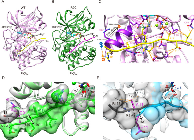
Figure 1.

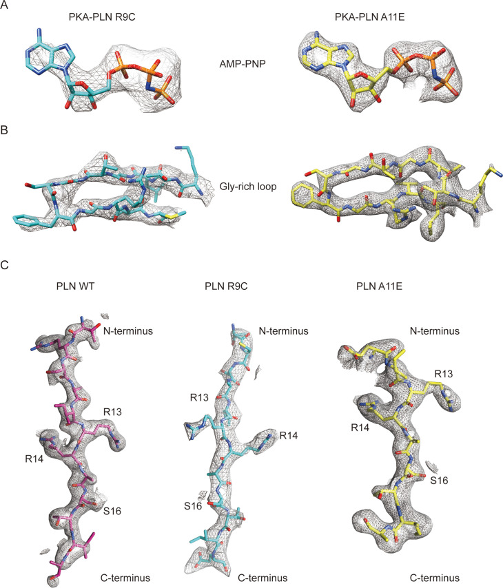
Figure 1—figure supplement 1. Electron density maps.

Figure 1—figure supplement 1.

Density maps for the AMP-PNP region (A), PKAc Gly-rich loop region (B), and phospholamban (PLN) peptides (C) from the crystal structures of PKAc–PLNs.
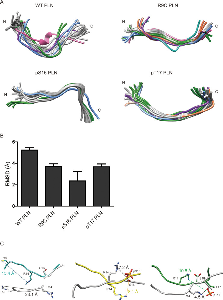
Figure 1—figure supplement 2. Nuclear magnetic resonance (NMR) structures of phospholamban (PLN)/ALN peptides in the absence of PKAc.

Figure 1—figure supplement 2.

(A) Ensemble cluster views of the superposition of the top 20 lowest energy structures of WT PLN, R9C PLN, pS16 PLN, and pT17 PLN. The top 7 ranked clusters are colored in gray, forest green, cornflower blue, purple, sandy brown, light sea green, and hot pink, respectively. (B) The root mean square deviation (RMSD) between the representative structures from each ensemble cluster. WT PLN clearly shows a higher RMSD than the other four peptides, suggesting its higher flexibility. (C) A comparison of the top structures of WT PLN (white) with the ones from R9C PLN (light sea green), pS16 PLN (yellow), and pT17 PLN PLN (forest green). S16 is used for the superposition.
Figure 1—figure supplement 3. Sequence alignment between phospholamban (PLN) and ALN.

Figure 1—figure supplement 3.

Identically conserved residues are colored in green, and weakly similar residues are coloured in grey. The protein kinase A (PKA)/calmodulin-dependent protein kinase II (CaMKII) phosphorylation site is colored in yellow and orange, respectively.
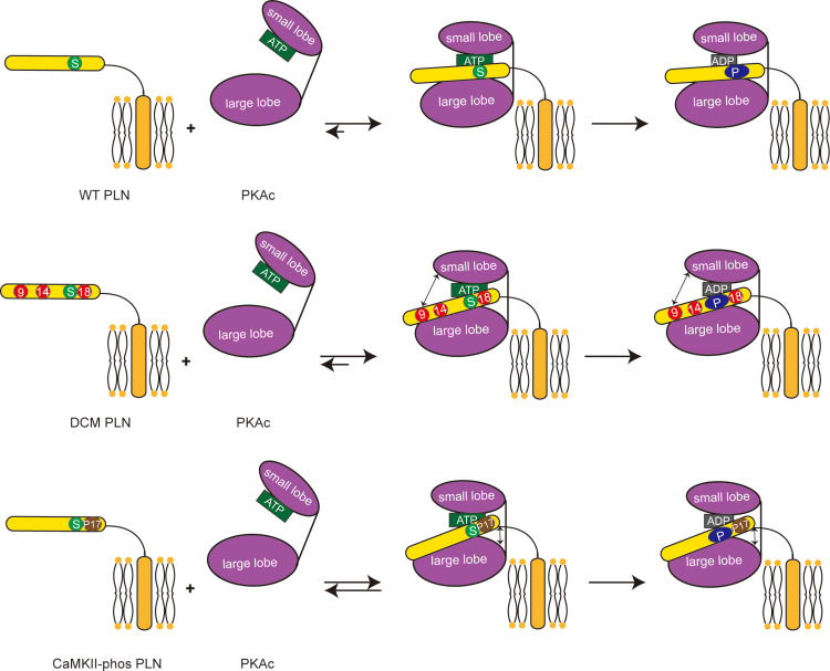
Figure 1—figure supplement 4. Schematic representation showing the impacts of dilated cardiomyopathy (DCM) mutations and calmodulin-dependent protein kinase II (CaMKII) phosphorylation on the phosphorylation of phospholamban (PLN) by protein kinase A (PKA).

Figure 1—figure supplement 4.

Both DCM mutations at the positions 9, 14, and 18 (red circles) and the CaMKII phosphorylation (brown circle) reduce the interaction between PLN (yellow) and PKAc (purple), and subsequently decrease the phosphorylation level of PLN.