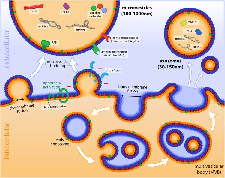
FIGURE 1.
Schematic illustration of microvesicles and endosomes and their formation. Important cargo molecules and markers of microvesicles and exosomes are shown in the lumen of EVs in the extracellular environment. The budding process of microvesicles by cis-membrane fusion is indicated (with the cytoplasmic sides of the membranes getting in first contact) as opposed to the trans-membrane fusion event of endocytosis. The exposure of tissue factor, as well as negatively charged PS (after flipping from the inner leaflet to the outside by cellular activation or apoptosis) is depicted on the budding microvesicle. The lower panel of the figure illustrates the process of endocytosis and the formation of intraluminal vesicles that are found in late endosomes (multivesicular bodies, MVBs) and how these vesicles are released as exosomes, when MVBs fuse with the plasma membrane.

