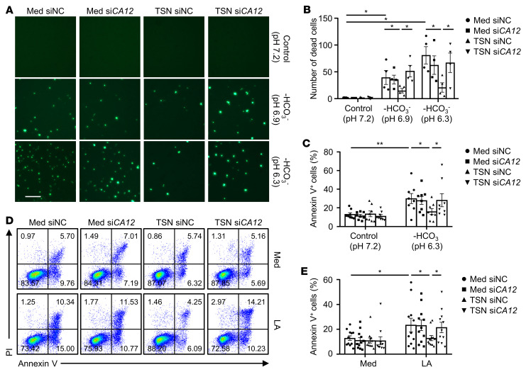
Figure 4. CA12 mediates the survival of monocytes and macrophages in acidic microenvironments.
(A–E) CD14+ cells were purified from the peripheral blood of healthy donors. Cells were transfected with siNC or siCA12 and then treated with or without HepG2 TSN for 48 hours before being exposed to pH 7.2 control medium (Med), pH 6.9 HCO3–-free medium, pH 6.3 HCO3–-free medium, or L-lactic acid (LA) (20 mM) for another 48 hours. (A and B) Dead cells were stained with SYTOX Green and analyzed by fluorescence microscopy (n = 4). One out of five representative graphs is shown in A. Scale bar: 50 μm. (C–E) Apoptosis of the cells was analyzed by flow cytometry. n = 8 (C); n = 11 (D and E). Results shown in B, C, and E are represented as mean ± SEM. P values were obtained by 1-way ANOVA. *P < 0.05; **P < 0.01.

