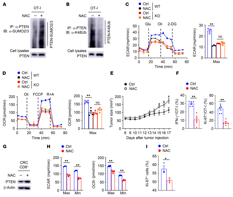
Figure 7. ROS involve SENP7-dependent CD8+ T cell metabolism and function.
(A and B) PTEN SUMOylation (A) and ubiquitination (B) assays using WT OT-I cells stimulated with anti-CD3 and anti-CD28 antibodies plus NAC for 2 hours. (C and D) ECAR (C) and OCR (D) of WT and KO OT-I cells stimulated with anti-CD3 and anti-CD28 antibodies plus NAC for 6 hours. (E) Tumor growth of MC38-OVA tumor–bearing WT mice (day 6 after tumor cell inoculation) injected i.v. with WT OT-I cells stimulated with anti-CD3 and anti-CD28 antibodies plus 10 mM NAC for 8 hours in vitro (control: n = 8; NAC: n = 9). (F) Flow cytometric analysis of the frequency of IFN-γ–producing and Ki-67+ OT-I cells in the tumors of mice from E (day 17 after tumor injection, n = 6). (G) Immunoblot analysis of the indicated proteins in CD8+ T cells from CRC samples incubated in vitro for 2 hours in complete media containing 10 mM NAC. (H) ECAR and OCR of CD8+ T cells from CRC samples incubated in vitro for 2 hours in complete media containing 10 mM NAC. (I) Flow cytometric analysis of the frequency of Ki-67+ CD8+ T cells from CRC samples incubated in vitro for 6 hours in complete media containing 10 mM NAC (n = 3). Data are representative of 3 independent experiments and presented as the mean ± SEM. *P < 0.05 and **P < 0.01, by 1-way ANOVA with Tukey’s multiple-comparison test (C and D) and 2-tailed Student’s t test (E, F, H, and I).

