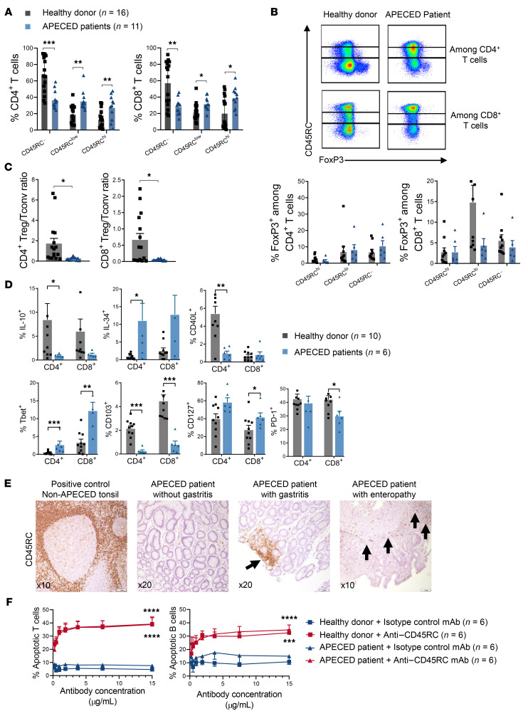
Figure 8. CD45RC expression is increased in peripheral blood T cells and autoimmune tissue lesions of APECED patients.
(A) PBMCs of APECED patients (n = 11) and healthy donors (n = 16) were stained for flow cytometry analysis showing the expression of CD45RC on CD4+ (left) and CD8+ (right) T cells; t test: *P < 0.05, **P < 0.01, ***P < 0.001. (B) Expression of FOXP3 and CD45RC on CD4+ (top line) and CD8+ T cells (bottom line) from APECED patients and healthy donors. (C) Ratio of FOXP3+ Tregs versus CD45RChi Tconv cells in healthy donors (n = 16) versus APECED patients (n = 11); t test: *P < 0.05. (D) Expression of IL-10, IL-34, CD40L, Tbet, CD103, CD127, and PD-1 by CD4+ and CD8+ T cells from healthy donors and APECED patients; t test: *P < 0.05, **P < 0.01, ***P < 0.001. (E) Representative immunohistochemical staining of CD45RC with an anti–human CD45RC mAb in stomach and small intestine paraffin-embedded tissue from 2 APECED patients with autoimmune gastritis and enteropathy (arrows) compared with stomach tissue of an APECED patient without autoimmune gastritis. Non-APECED human tonsil biopsy tissue was used as positive control. (F) Proportion of apoptotic CD45RAhi T cells induced after a 3-hour in vitro incubation of PBMCs, from healthy donors (n = 6) or APECED patients (n = 6) with the anti-CD45RC or isotype control mAbs. One-way ANOVA repeated measures, Bonferroni’s post hoc test: ***P < 0.001, ****P < 0.0001.

