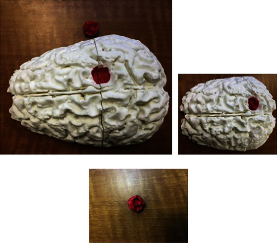
Figure 2.

Postprinting result. (a, b) The printed brain can be combined with the tumor print (red) in order to establish the relationships of the adjacent anatomic structures. (c) The tumor can be painted to determine the separation from the brain parenchyma. This is a cost-effective procedure that can help to improve the three-dimensional visualization of the brain tumors to improve the management [71]. Used with permission from Elsevier.
