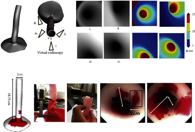
Figure 5.

(a) Visualization of 3D depth maps models of the esophagus. Techniques to determine the localization, length, and depth of the Barret's lesions through the endoscopy camera. (b) 3D printed model of the esophagus that shows the measurements for the lesions (C and M) and also the endoscopy video frames are shown [163]. Used with permission from Elsevier.
