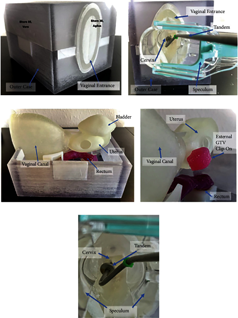
Figure 7.

(a) Closed 3D printed model. (b) The speculum entering the vaginal canal. (c) 3D printed intrapelvic organs and its anatomical position. (d) Presence of 3D printed gross tumor attached to the uterine body. (e) Tandem used in brachytherapy procedures is inserted through the speculum and placed inside the cervix and uterine canal. These phantom models help in teaching physicians the process of intracavitary procedures in cervical cancers [201]. Used with permission from Elsevier.
