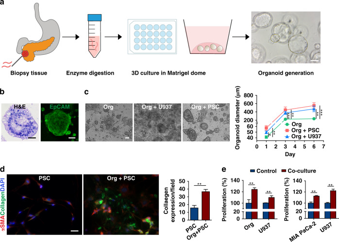
Fig. 1. Patient-derived organoid (PDOs) propagation, characterization, and bidirectional interactions with stromal cells in welled plates.
a Schematic describing the workflow used to generate PDOs from biopsy samples via fine-needle biopsy (FNB). PDOs (generated from the biopsy sample of one patient) formed sphere-shaped cell clusters in 3D Matrigel culture. Scale bar, 100 µm. b Characterization of the isolated PDOs by H&E and EpCAM staining. Scale bar, 100 µm. c PDOs cocultured with PSCs and U937 monocytes showed an increased average diameter (±SEM, n = 3) over a 6 day culture period in welled plates. Scale bar, 100 µm. d Coculture of the organoids with PSCs significantly increased collagen deposition (n = 3 wells, expression quantified from ten fields). Scale bar, 20 µm. E Bidirectional increase in the proliferation of neoplastic epithelial cells (PDO and MIA PaCa-2) and U937 monocytes in a transwell culture assessed by a cell-counting kit assay (n = 5, CCK8). *p < 0.05, **p < 0.01, ***p < 0.001

