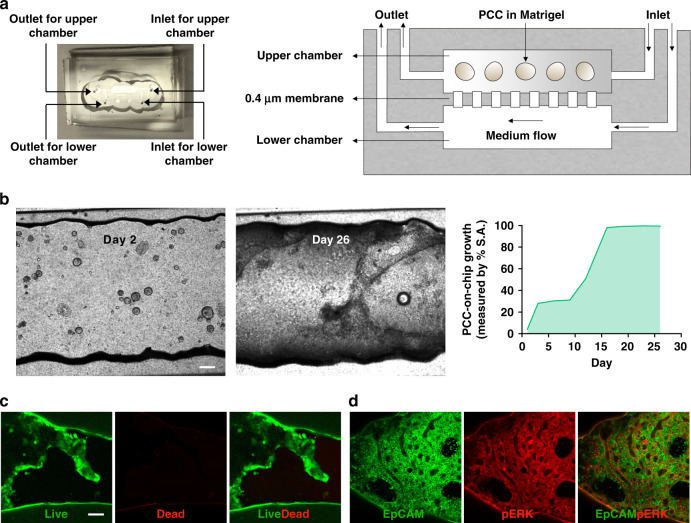
Fig. 3. A two-chamber microfluidic chip for the seeding and growing of PDOs.
a Image and schematic of the multichamber microfluidic device. The chip consists of two chambers separated by a 0.4 µm porous membrane. Cells are installed in the upper chamber through the inlet. Perfusion of the cell culture medium through the lower chamber maintains cell viability. b Top view images of the cell-laden chip. PDOs were cultured for 26 days with continuous perfusion of the medium. After 1 week, the organoids lost their 3D spherical shapes and spread two-dimensionally to occupy the inner surface area (S.A.) of the chip (right panel). On Day 26, c viability and d pERK expression were assessed in the PCCs to determine survival in the organ-chip device. Scale bar, 200 µm

