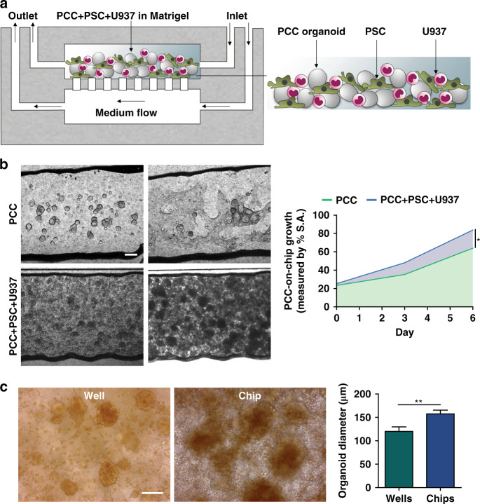
Fig. 4. Growth of the primary cancer cells (PCC) with stromal cells in chips.
a Schematic of PCC + stromal cell seeding on the microfluidic device (n = 3 chips). b PCCs grew to significantly higher density when cocultured with stromal cells and occupied more space than PCCs in PCC monoculture in chips, indicating ongoing bidirectional proliferation. Scale bar, 200 µm. The area-filled graph shows the PCC growth dynamics on the chip’s inner surface area (S.A.). c Organoids that grew on a chip had a significantly greater diameter than the organoids grown in a welled plate. Images were acquired from organoids cultured in three wells/chips and quantified from ten fields using ImageJ software to determine the average organoid diameter ± SEM. Scale bar, 100 µm. *p < 0.05, **p < 0.01

