Figure 7.
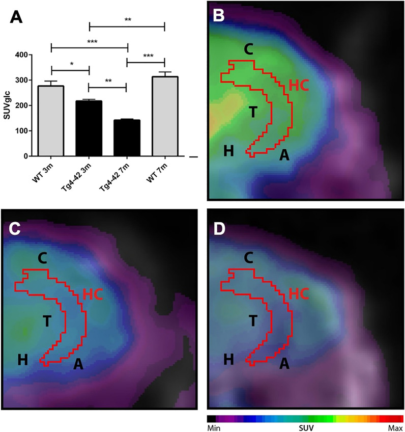
18F-FDG-PET shows decreased metabolic activity in the hippocampus of Tg4-42 mice. (A) Quantification of 18F-FDG uptake in the hippocampus. 18F-FDG-uptake in the hippocampus was significantly reduced in 3m and 7m Tg4-42 mice compared to same-aged WT animals. Hypometabolism in Tg4-42 increases age-dependently. (B) Fused 18F-FDG-PET/MRI of a WT mouse in coronal view. (C) Fused 18F-FDG-PET/MRI of a 3-month-old Tg4-42 mouse in coronal view with distinctly lower FDG uptake compared to the WT mouse. (D) Fused 18F-FDG-PET/MRI of a 7-month-old Tg4-42 mouse in coronal view with distinctly lower FDG uptake compared to WT and 3-month-old Tg4-42 mice. One-way-ANOVA; ***p < 0.001; **p < 0.01; *p < 0.05; WT wild-type; m months. A Amygdala, C Cortex, H Hypothalamus, Hc Hippocampus, T Thalamus.

