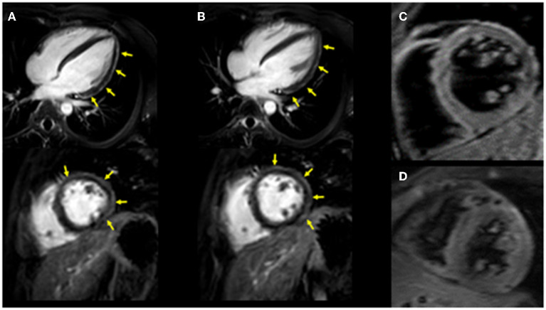
Figure 1.
Cardiac MRI imaging. Diffuse late gadolinium enhancement at the epicardium was observed in both images. (A) Images obtained 3 years ago when he suffered from his previous myocarditis (top: long-axis view, bottom: short-axis view). (B) Images obtained during the current myocarditis episode associated with coronavirus disease-2019 (COVID-19) messenger RNA (mRNA) vaccination (top: long-axis view, bottom: short-axis view). T2-weighted MR images. (C) Image obtained 3 years ago when he suffered from his previous myocarditis episode. (D) Image obtained during the current myocarditis episode associated with COVID-19 mRNA vaccination.

