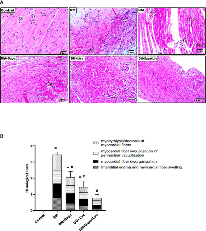
Figure 2.
(A) Representative photomicrographs of H&E-stained cardiac sections from different experimental groups. Normal control rats showed normal histological structure of the cardiac muscle diabetic group demonstrating disarray of the cardiac myocytes with myocardial fiber disorganization (curved arrow), myocardial fiber necrosis with chronic inflammatory cells in the subpericardium (astrix), apoptotic myocyte with hypereosinophilic cytoplasm, pyknotic nuclei (crossed arrow), increased intermyocyte (thick arrows), and perivascular chronic inflammatory cells (zigzag arrows) marked interstitial edema. Treatment groups (DM+Dapa) and (DM+Lira) showed less structural injury. Combination therapy group (DM+Dapa+Lira) showed almost restoration of normal cardiac structure. ×400 bar 25. (B) A graph showing histological score for cardiomyopathy. Data are expressed as mean ± SEM (n = 6). *p < 0.05 versus control and # p < 0.05 versus diabetic group. Dapa, dapagliflozin; Lira, liraglutide.

