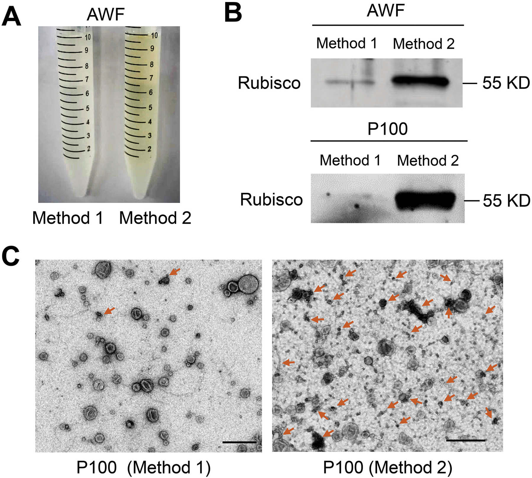
Figure 2. The detached leaves protocol (Method 1) for AWF isolation is better than the whole plant protocol (Method 2) in Arabidopsis.

(A) Comparison of the color of AWF isolated by Method 1 and Method 2. The same amount of plants (50 plants) was used for both methods. (B) Detection of Rubisco protein in both AWF and their P100 EV fraction by Western blot using Rubisco antibody, protein size is indicated by KD. To perform the Western blot of AWF samples, equivalent amounts of AWF (10 μl) collected by both methods in (A) were used. To perform the Western blot of EV samples, all AWF collected in (A) was centrifuged at 100, 000 × g to get P100 fractions. Both P100 pellets were resuspended in 100 μl infiltration buffer, and 10 μl of this suspension was used for the Western blot. (C) Representative transmission electron microscopy (TEM) images of P100 fraction isolated from AWF collected by Method 1 and Method 2. Non-vesicle structures marked by arrows. Scale bars, 500 nm.
