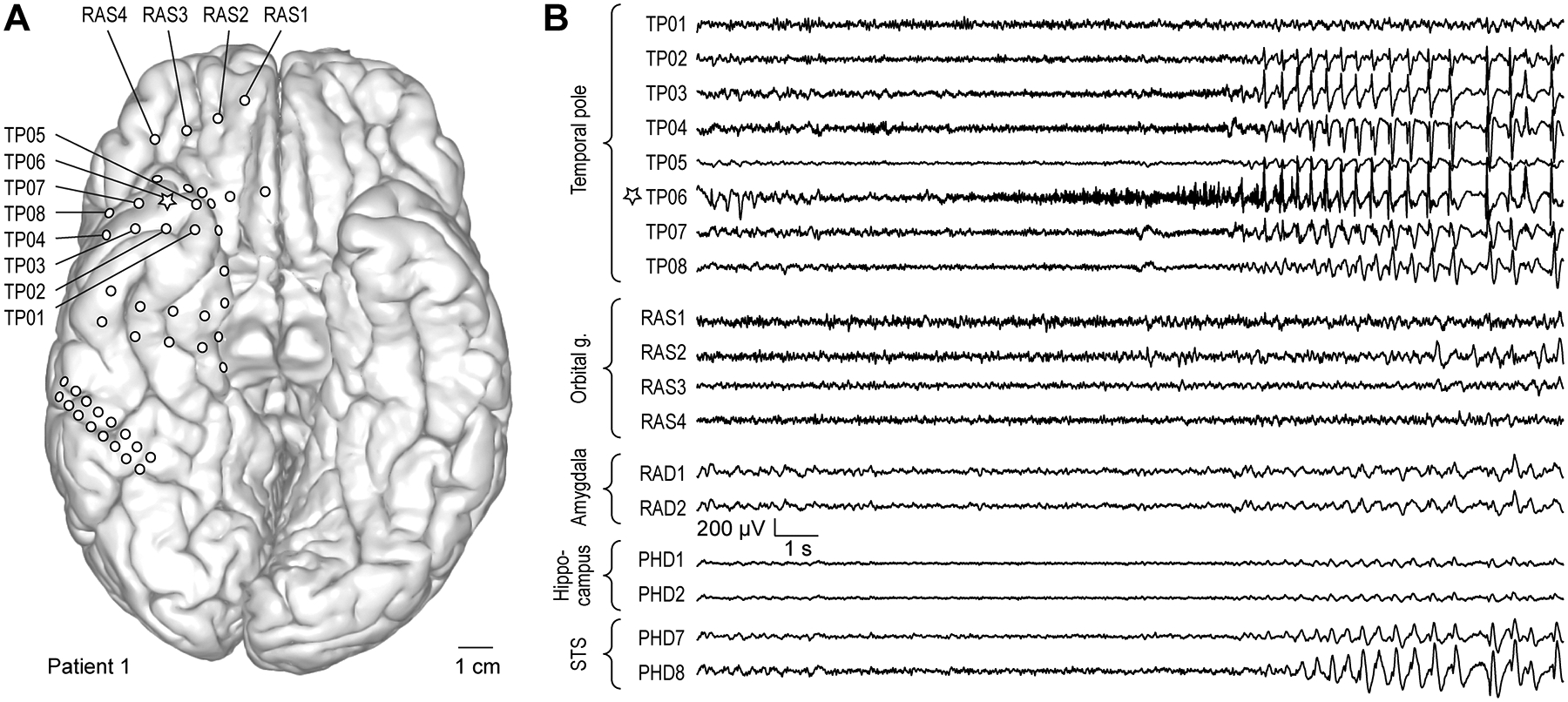
Figure 2:

Exemplary seizure. A: Anatomical reconstruction of ventral brain surface demonstrating location of implanted epicortical electrode arrays in a representative patient (#1). Star denotes the recording site where seizure onset was observed (TP06). B: Exemplary recordings from epicortical electrodes implanted over TP and orbital gyrus, and depth electrodes implanted in the amygdala, hippocampus and superior temporal sulcus (STS).
