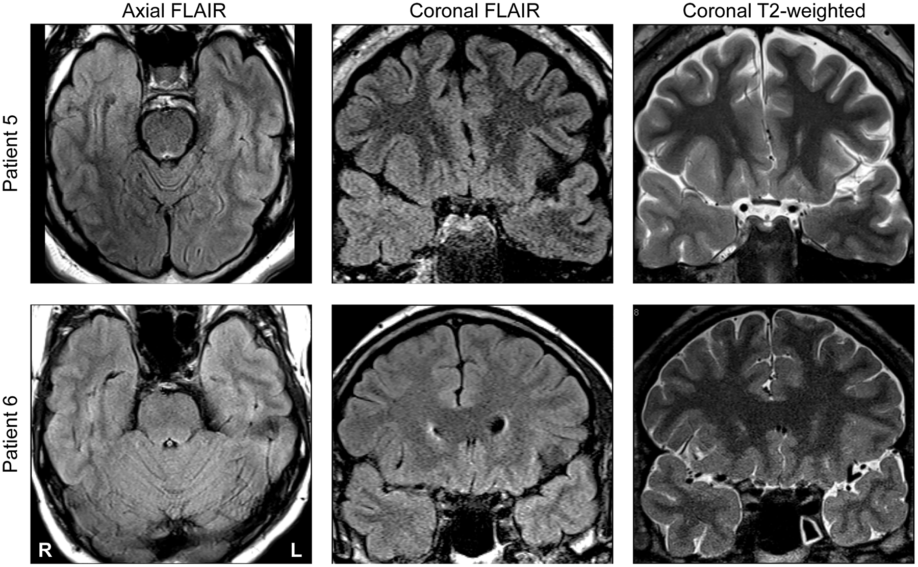
Figure 3:

Neuroimaging findings in two representative patients (#5 and #6; top and bottom row, respectively). Axial FLAIR, coronal FLAIR and coronal T2-weighted scans are shown in left, middle and right columns, respectively. In each patient, there is clear atrophy and FLAIR signal abnormality of temporopolar cortex manifest on MR imaging.
