
Figure 2. Fundus images from a patient from Family B with EFEMP1 variant p.Ter494Glnext*29 (B-V:5).
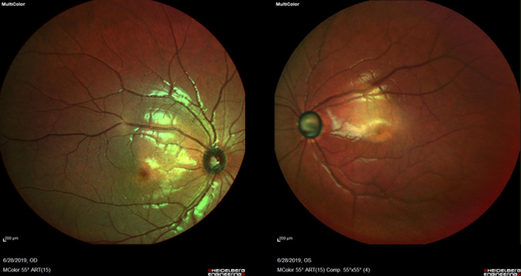
The image shows damage to the optic nerve but no evidence of subretinal deposits (drusen) characteristic of Malattia Leventinese/Doyne Honeycomb dystrophy. This non-mydriatic fundus photo was obtained using Heidelberg Spectralis® MultiColor Imaging (Heidelberg Engineering, Heidelberg, Germany).