Figure 1.
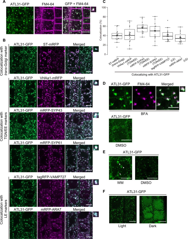
ATL31 localizes to the plasma membrane and endosomal compartments. A, Representative confocal images of FM4–64-stained Arabidopsis root epidermal cells expressing ATL31-GFP. The root was stained with 2-µM FM4–64 for 2 min and washed with medium without dye. The photograph was taken 18 min after staining. Bar = 10 µm. B, Representative confocal images of Arabidopsis root epidermal cells co-expressing ATL31-GFP with ST-mRFP (trans-Golgi), mRFP-SYP43 (TGN/EE), mRFP-SYP61 (TGN/EE), VHAa1-mRFP (TGN/EE), mRFP-ARA7 (LE), and tagRFP-VAMP727 (LE; F1 generation). Small panels on the right show enlarged views of the dot-like structures. Bars = 10 µm. C, Quantification of the data in (B). Percentage of dot-like structures of ATL31-GFP colocalizing with the indicated markers. The center–center distances of the nearest dot-like structures were calculated using the ImageJ plugin DiAna, and endosomes at a distance closer than 0.2 µm (approximate theoretical resolution limit of the confocal microscope) were considered to be colocalizing. n = 15 cell slices from five roots were analyzed. In the box plots, center line, median; box limits, lower and upper quartiles; +, mean; dots, individual data points; whiskers, highest and lowest data points (the whiskers extend to data points that are less than 1.5× interquartile range (IQR) away from the 1st and 3rd quartile.). D, Representative confocal images of Arabidopsis root epidermal cells expressing ATL31-GFP treated with 50-µM BFA or DMSO for 30 min. Cells were stained with 5-µM FM4–64 15 min before BFA treatment. The small panel on the right shows and enlarged view of BFA bodies. Bars = 10 µm. E, Representative confocal images of Arabidopsis root epidermal cells expressing ATL31-GFP treated with 33-µM WM or DMSO for 1 h. Arrowheads indicate the ring-like structures representing vacuolized late endosomal compartments. Bars = 10 µm. F, Representative confocal images of Arabidopsis root epidermal cells expressing ATL31-GFP after 18 h of light or dark treatment. Bars = 10 µm.

