Figure 7.
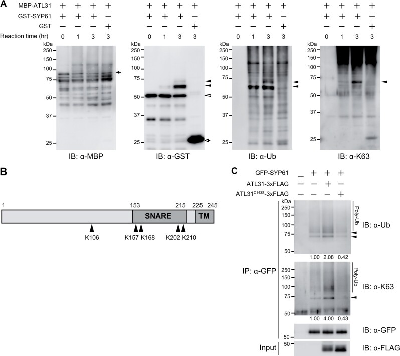
ATL31 ubiquitinates SYP61 in vitro and in planta. A, In vitro ubiquitination assay of SYP61 by ATL31. GST-SYP61 or GST was incubated with MBP-ATL31 in the presence of ubiquitin, E1, E2, and ATP for the indicated time. Immunoblot analysis was performed with anti-MBP, anti-GST, anti-ubiquitin (FK2), and anti-K63-linked ubiquitin (Apu3) antibodies. Closed arrow, MBP-ATL31 (81 kDa); open arrow, GST (28 kDa); open arrowhead, GST-SYP61 (53 kDa); closed arrowheads, ubiquitinated SYP61. B, Schematic diagram of the primary structure of SYP61 protein. Arrowheads indicate the ATL31-catalyzed ubiquitination sites of SYP61, as determined by mass spectrometry analysis following an in vitro ubiquitination assay as in A. Numbers indicate the amino acid positions. SNARE, SNARE domain; TM, transmembrane domain. The original data are shown in Supplemental Table S1. C, Immunoblot analysis to examine SYP61 ubiquitination by ATL31 in plants. GFP-SYP61 (56 kDa) was transiently expressed alone or with ATL31-3xFLAG or ATL31C143S-3xFLAG (45 kDa) in N. benthamiana leaves. Extracted proteins were immunoprecipitated with anti-GFP antibody beads and detected with anti-GFP, anti-ubiquitin (FK2), and anti-K63-linked ubiquitin (Apu3) antibodies. Arrowheads indicate ubiquitinated GFP-SYP61. The numbers show the relative intensities of the di-Ub bands (the bands indicated by the lower arrowhead in the anti-ubiquitin blot) normalized by the band intensity of GFP-SYP61.

