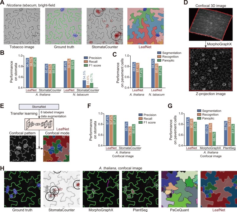
Figure 5.
Extension of LeafNet to analyze species with similar morphology as well as confocal images. A, Representative example of LeafNet results for a bright-field image of N. tabacum. Raw image, ground truth labeling, StomataCounter results, and LeafNet results are shown from left to right. B, Performance of LeafNet for stoma detection in A. thaliana and N. tabacum bright-field images compared with StomataCounter. C, Performance of LeafNet for segmenting pavement cells in A. thaliana and N. tabacum bright-field images using three different metrics. D, Representative example of preprocessing a confocal 3D image to a z-projection image using MorphoGraphX. E, Extension of LeafNet to analyze z-projection image using transfer learning based on limited number of newly labeled data to identify stomata with its confocal mode. F, Performance of LeafNet for detecting stomata in A. thaliana confocal images compared with StomataCounter. G, Performance of LeafNet for pavement cell segmentation in A. thaliana confocal images compared with MorphoGraphX and PlantSeg for three different metrics. H, Representative outputs of five programs using confocal images. StomataCounter, PlantSeg, PaCeQuant, and LeafNet use max intensity z-projection images as input, while MorphoGraphX takes 3D image stacks as input.

