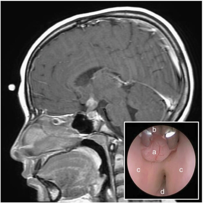
Fig. 2.
Sagittal post-contrast MRI of the brain demonstrating a hypothalamic and infundibular mass in a 10-year-old female presenting with diabetes insipidus. Given normal serum and CSF tumor markers, a right frontal endoscopic tumor biopsy was performed which confirmed pure germinoma. Endoscopic view of the third ventricle (inset) revealing the exophytic tumor mass (a), 1 mm endoscopic biopsy forceps (b), and the bilateral hypothalami (c).

